A pig looking out of their barren crate in a factory farm.

The zoonotic disease burden in Australia.

The global practice of confining and slaughtering animals, both farmed and wild, inflicts severe cruelty on billions of sensitive individuals annually – it also provides the perfect conditions for infectious diseases to emerge and spread.

Animals Australia

Animals Australia team

Last updated May 5, 2020

Zoonotic diseases are diseases that arise in animals, jump the species barrier to humans, and then spread via human-to-human transmission.

Many, and arguably all, zoonotic diseases arise because of the ways humans treat animals – both directly, by keeping them in factory farms and ‘wet markets’, and indirectly, through actions like habitat destruction which increases contact between wildlife, people, and farmed animals.

From bird and swine flu to SARS, MERS, anthrax, and others, there is a strong case that the treatment of animals in our broken food systems is a major factor in the spread of new diseases on a global scale.

Bird Flu

Avian influenza viruses may be introduced to Australian commercial poultry farms through contact with wild birds and ducks (the natural reservoirs). The viruses then circulate within poultry populations, mutating on-farm and occasionally becoming highly pathogenic due to the large numbers of birds forced to live within a shed and potentially amongst rows of sheds.

From there, the virus may be spread off the farm by workers; when the poultry are transported in trucks to a slaughterhouse; in the poultry’s waste, which may be sprayed across land or dumped in a water body; by wild birds, flies and other insects; and by high-volume fans that are necessary to ventilate the densely packed sheds.[1]

A schematic representation (not to scale) of multiple potential pathways for exposure to, and transfer of, pathogens in an animal farming operation:[1]

This image contains content which some may find confronting

illustration of how pathogens could be distributed around a chicken farm
Compromises to biosecurity include: (1) workers lacking protective clothing or opportunities for personal hygiene or decontamination on-site; (2) inadequate management of animal biosolids, often applied to land without treatment; (3) flies and other insects that carry pathogens in and out of facilities through ventilation systems and small openings; (4) ventilation with high-volume fans resulting in considerable movement of materials into the external environment; and (5) transporting animals in open trucks or containers to the farm or for processing.
Image credit: Salvador Saenz

At least seven highly pathogenic avian influenza (HPAI) outbreaks and fifteen low pathogenic avian influenza (LPAI) outbreaks have occurred in Australian poultry farms since 1976.[2][3] Poor conditions on-farm were identified in at least three of the seven HPAI outbreaks.

Outbreaks of Highly Pathogenic Avian Influenza in Australian poultry farms:

  • 2024: In Meredith (Vic), hundreds of thousands of egg-laying hens were killed at a battery cage facility after the detection of avian influenza after reports of deaths at the property.
  • 2013: In Young (NSW), an outbreak of H7N2 occurred on an egg farm housing over 400,000 layer hens across 8 free range and 6 caged sheds.[4] The outbreak spread to another egg farm 35km away, which housed 40,000 layer hens. All birds were destroyed.[5]
  • 2012: In Maitland (NSW), an outbreak of H7N7 occurred on an egg farm and some 50,000 layer hens were destroyed.[6]

  • 1997: In Tamworth (NSW), an outbreak of H7N4 occurred on two broiler chicken breeding farms and an emu farm. The first farm housed 128,000 chickens across 15 sheds. The second farm had 32,000 chickens across 6 sheds. Subsequent surveillance of nearby properties detected the same virus in a flock of 3-month-old emus on a contract grower farm, though they were asymptomatic. Following confirmation of HPAI, all three farms were ‘depopulated’; a total of 310,565 chickens, 261 emus, over 1.2 million fertile chicken eggs and 261 emu eggs were destroyed. Transmission of the virus among the chicken farms was attributed to a dead bird pick-up truck and worker.[7]
  • 1994: In Lowood (Qld), an outbreak of H7N3 occurred on an egg farm housing approximately 22,000 layer hens aged from 6 to 40 weeks. The source of the outbreak was suspected to be river water heavily populated with wild ducks due to the drought, though this was never proven.[8]
  • 1992: Near Bendigo (Vic), an outbreak of H7N3 occurred on a ‘meat’ chicken breeding farm. The source of the outbreak was believed to be a neighbouring, poorly managed duck farm housing 5,700 ‘meat’ ducks. The owner of the duck farm worked periodically on the affected chicken farm and may have inadvertently spread the virus to (or from) the chickens. All of the birds were killed.[9]
  • 1985: Near Bendigo (Vic), an outbreak of H7N7 occurred on a chicken farm housing 120,000 chickens across 12 sheds. The chickens comprised ‘meat’ chickens and breeders, egg-laying hens and started pullets. The property also incorporated a small abattoir. The farm had a continuous history of various low-grade disease problems. The source was suspected to be wild ducks using a lake on the farm, though this was never proven.[10]
  • 1976: In Keysborough (Vic), an outbreak of H7N7 occurred on a combined ‘meat’ chicken and egg-laying hen farm. Subsequent surveillance identified an H7 virus present in the duck farm opposite, and this was believed to be the source of the chicken farm outbreak. The duck farm was in a disgraceful condition with dilapidated sheds and poor oversight of the animals.[11]

This image contains content which some may find confronting

A young 'broiler' chicken suffering in a poultry farm.
Chicken sheds can confine tens of thousands of individuals. Through selective breeding and feed manipulation, chickens have been 'modified' to grow so fast that they reach 'slaughter weight' when they're only 5-8 weeks old. Many of these young birds will be crippled by their own weight and unable to get up off filthy factory farm floors.

Swine flu

The Swine Influenza H1N1 pandemic of 2009 is estimated to have caused as many as 575,400 human deaths worldwide.[12] The virus emerged in California in April among people who had direct contact with pigs. By May, it had infected people in Australia, and by August, it had caused outbreaks in three commercial piggeries across NSW[13], Victoria [14] and Queensland[15]. The H1N1 pandemic virus has been circulating in commercial Australian piggeries with sporadic outbreaks ever since.[16]

This image contains content which some may find confronting

pigs in transport truck
There are over 1,000 pig farms in Australia. It's common for piglets bred into this industry have their teeth and tails cut off – without any pain relief. Cutting off body parts is the industry’s ‘solution’ to behaviours that are associated with their confinement, rather than giving these social, clever animals more space and enrichment.

This trend of human influenza pandemic viruses entering pig farms is a global one, and not limited to the 2009 H1N1 pandemic. Genetic analysis of tissue samples collected from sick pigs at commercial piggeries in Western Australia and Queensland revealed the transmission of multiple H1N1 and H3N2 influenza A viruses, including some derived from the 1977, 1995, 2003 and 2009 human outbreaks.[17]

Novel swine influenza viruses have also been detected in Australian people and piggeries. In 2012, an outbreak of respiratory illness and deaths occurred in a piggery near Perth and was accompanied by a mild respiratory illness reported by several of the workers. Analysis of the pig samples taken revealed an unidentified swine influenza A virus containing genes of human origin.[18] More recently, in September 2018, a 15-year-old girl from a semi-rural area in South Australia became infected with Swine Influenza variant H3N2 after exposure to pigs at a ‘livestock’ exhibition. The virus was detected during routine screening of influenza-positive samples.[19][20]

Q Fever

Q Fever is caused by the highly pathogenic bacteria Coxiella burnetti, and symptoms can vary from a mild influenza-like illness to a fatal acute condition. The bacteria are found in birthing fluids, milk, urine and faeces, and can become airborne, spreading over wide areas in the wind and in air conditioning systems. Cattle, sheep and goats are the main reservoirs, and human infections typically result from slaughtering animals or assisting with animal births.

A study of Q Fever cases across a decade in South Australia found that the disease primarily affected men aged 21-40 years old, with most cases occurring through occupation (livestock farmers or slaughterhouse workers) or residence (living in a suburb with a slaughterhouse).[21]

Q Fever was first described in Australian slaughterhouse workers in the 1930s, though it occurs across the globe. Since 1990, there have been 300-800 confirmed human cases of Q Fever in Australia each year.[22] However, seroprevalence studies reveal 30-70% of slaughterhouse workers have been exposed to the bacteria, suggesting the burden of Q Fever is much larger than the number of annual case notifications.[23]

This image contains content which some may find confronting

Q Fever
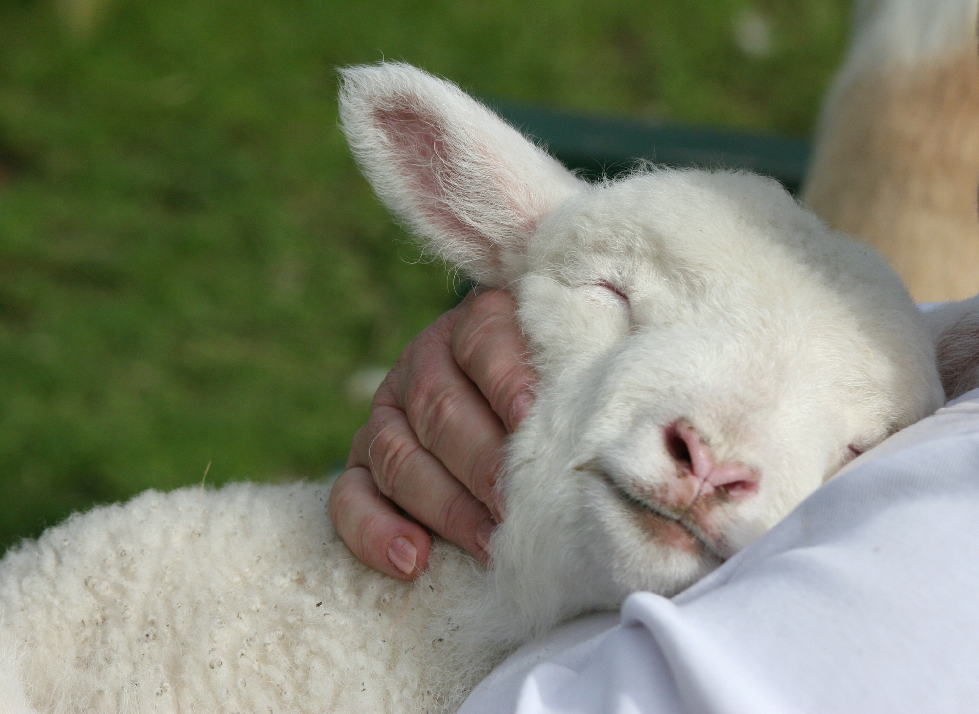
Millions of lambs are bred to be slaughtered each year in Australia. This lamb, only minutes old, was born on the cold, wet concrete of a saleyard floor. Despite his mother having been in labour during transport, and having just given birth in the holding pens, she was next on her way to slaughter.

Outbreaks of Q Fever in Australia, including cases where the bacteria has become airborne, have been traced to saleyards>[24], intensive dairy farms[25] and slaughterhouses, including facilities that process cattle placentas[26].

Q fever has the potential to cause large epidemics, as demonstrated in the Netherlands from 2007-2010. Some thirty intensive dairy goat and sheep farms became infected, and more than 4,000 people were confirmed Q Fever cases (though the number of people infected was likely over 40,000). The disease spread to people living within 5 kilometers of an infected farm, and to people visiting and interacting with the young goats and lambs.[27][28]

Anthrax

Anthrax is an ancient disease caused by the bacteria Bacillus anthracis, which lives in soil and primarily infects herbivores like cattle, sheep, goats and deer. People become sick through contact with infected animals, and the type of illness a person develops depends on how the anthrax spores enter the body. Anthrax can be fatal if not treated with antibiotics and is notoriously known as a potential bioweapon.

Anthrax in Australian farmed animals was first recorded in 1847 at Leppington (NSW), spreading through cattle and sheep stock routes to Victoria, where it was first recorded around the Warrnambool area in 1886. From 1900 to the 1920s, there were approximately 80 outbreaks in NSW and 40 outbreaks in Victoria. Cases continued to be reported in NSW, with around 347 outbreaks from 1930-1962. Major outbreaks in Victorian cattle and sheep have occurred across 27 farms in the Yarrawonga/Shepparton area during 1968; across 83 dairy farms in the Stanhope/Tatura area during 1997; and across 10 farms in the Goulburn Valley area during 2007. Another large outbreak occurred simultaneously in NSW in 2007, involving 10 dairy farms across the Hunter Valley.[29]

This image contains content which some may find confronting

Anthrax
There are well over 20 million cattle in Australia used for dairy, for their meat, or both. Each one of them needs land to graze on and/or supplementary feed, taking up land across Australia that was once habitat to native animals.

More recently, anthrax outbreaks on sheep or cattle farms in NSW have occurred in the Parkes area (2015), Rankins Springs district (2016), Cumnock district of the Central Tablelands (2016 and 2018), Forbes district (2017), the Central West region (two outbreaks in 2018), and the Nyngan district (2019).[30] There have also been reported anthrax cases on sheep farms around the Swan Hill area of Victoria in 2017 (5 farms) and 2018 (two properties).[31]

Food-borne disease

Two of the most common and costly gastrointestinal diseases in Australia — Campylobacteriosis and Salmonellosis[32],[33] — are primarily caused by eating contaminated animal-based foods. Chicken meat has been implicated in 85% of foodborne Campylobacter outbreaks where a source was identified.[34] Thousands of non-typhoidal Salmonella cases from numerous outbreaks have been traced to eggs — a trend that is increasing.[35] Eggs have been implicated in 71% of Salmonella Typhimurium outbreaks where a source was identified. Furthermore, the burden of Salmonellosis in Australia is suspected to be seven times greater than the number of yearly case notifications.[36]

Scientific publications also reveal the routine detection of numerous pathogenic bacteria and parasites in farms, saleyards and slaughterhouses across the country.[37] From Listeria monocytogenes to Escherichia coli, Yersinia enterocolitica, Clostridium, Bacillus cereus, Cryptosporidium and Toxoplasma gondii — all of them (and many more!) can be found in places where Australian farm animals are raised and killed. Other studies reveal the presence of these harmful pathogens in animal-based foods available from Australian shops.[38]

The widespread use of antibiotics on farms to combat these bacteria is contributing to the evolution of antimicrobial resistance and ‘superbugs’ that pose significant risks to public human health.

This image contains content which some may find confronting

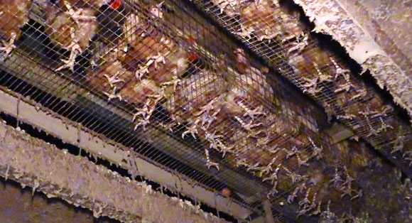
The feet of crowded hens in cages poke through the wire floor.
In these cramped conditions, hens are forced to stand, sleep, and toilet in the same spot. Battery cages not only deprive hens of a life worth living, but they can also foster the spread of disease among confined birds.

Importantly, this is all to be expected because farmed animals are the hosts for many micro-organisms, which may live in their digestive systems or be found on their skin, hair, wool and feathers. The presence and concentration of many pathogenic micro-organisms are directly influenced by the animals’ living conditions. Stocking density, weather, poor handling practices as well as dirty farm and transportation conditions all contribute to the presence of harmful pathogens in farmed animals, while stress increases the concentration of pathogens in their faeces.[39][40]

These pathogens readily contaminate meat products through contact with the animals’ faeces and entrails as the carcass is being skinned or de-feathered, eviscerated and processed; from the slaughterhouse environment or workers along the slaughterhouse processing line.[41][42] Bacteria that contaminate milk may come from the lactating mother’s body (especially if she has developed mastitis), from faeces, contaminated feed, housing or water — and, importantly, some harmful bacteria can survive pasteurization.[43]

This image contains content which some may find confronting

Graph of campylobacteriosis salmonellosis
National notifiable diseases surveillance system annual notifications (1991 to 2019) of Campylobacteriosis and Salmonellosis in Australia.

Finally, Salmonella bacteria can colonize the inside of an egg while it is developing within a layer hen, or the shell surface when it is laid in a contaminated environment or moved through contaminated equipment. And though most eggs are washed prior to being sold, this does not reduce the concentration of bacteria living within pores along the eggshell surface.[44][45]

Contact between eggshell and faeces is difficult to avoid. Even a visually 'clean' egg may have residual faecal material on the surface.

Psittacosis

Psittacosis (also known as Ornithosis) is a lung infection that may be asymptomatic or cause pneumonia. Small epidemics have been reported worldwide for over 100 years, with some involving >700 cases and 20-30% fatality rates.[46] In Australia, there are up to 230 cases diagnosed each year, though this is expected to be a substantial underestimate of Psittacosis prevalence.[47][48] The disease is caused by inhaling the excrement or other aerosols generated by birds infected with Chlamydia psittaci bacteria.

This pathogen has been found in over 450 bird species[49], though recent evidence from Australia suggests that C. psittaci infection is ten times higher in captive birds than in wild birds.[50] Other Australian research suggests that wild birds entering the pet trade experience a stress-induced ‘shedding’ of various pathogens, meaning captive birds have a greater concentration of pathogenic bacteria and viruses in their faeces because they are stressed.[51]

This image contains content which some may find confronting

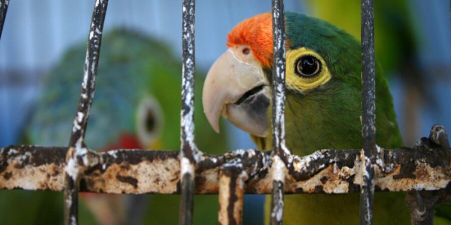
Colourful bird in a cage
Caging birds denies them the opportunity to express their desire to fly free, be social, raise their young in privacy, and engage in other natural behaviours.

Many psittacosis outbreaks have been traced to newly exported/imported sick birds within the wildlife trade. It was first demonstrated that Australian parrots in export shipments were harboring and spreading the disease in the early 1930s.[52] The export of native Australian birds is still a thriving trade — both legally and illegally.[53][54]

Other outbreaks in Australia have been traced to birds in pet shops[55], duck farms and slaughterhouses[56][57][58], a game bird processing facility[59] and, most recently, a thoroughbred horse in a veterinary clinic.[60]

Menangle Virus

Menangle Virus emerged during a single outbreak among intensively farmed pigs and piggery workers in Menangle (NSW) during 1997. This virus circulates naturally among flying foxes (fruit bats) and it is likely pigs became infected from exposure to bat urine or other bodily fluids. Infected pigs primarily suffered reproductive losses — stillborn and mummified piglets — while human infection resulted in a severe influenza-like illness.[64]

The piggery had 2,600 sows and comprised four sets of sheds, located immediately alongside a forested flying fox roosting area. Young females selected for breeding were housed in units 1 and 2, while units 3 and 4 were the farrowing areas and where sows were confined for the majority of their pregnancies. These units were closest to the bat roosting area. Between 8-24 pregnant sows were kept together throughout their pregnancies. Piglets were weaned at 18-24 days old and moved to ‘growing sheds’ either on-site or several hundred kilometers away in Trunkey Creek and Young. Each week, a group of 125 piglets were moved among the shed units.[64b]

This image contains content which some may find confronting

Satellite image of a piggery

Development Applications for intensive farms, such as the piggery above, currently require comprehensive environmental impact statements – in theory. As a matter of urgency, these applications ought to also require a thorough analysis of the local wildlife and, if any flying foxes or other potential viral hosts are detected, they must be denied in the interests of public health and safety.

Brucellosis

A recent global review of diseases at the wildlife-livestock interface ranked brucellosis among the top ten diseases.[65] Symptoms in people are typically non-specific and flu-like, sometimes including severe depression, and have an overall case fatality rate of 2%.

In Australia, brucellosis is caused by Brucella suis bacteria that live in wild pigs across Queensland and New South Wales.[66] Transmission to people and dogs occurs during pig hunting and eating uncooked wild pig meat, by direct contact with infected pig blood or bodily fluids, or by aerosol spread.[67] Infected pig hunting dogs can also pass the disease to breeders, handlers, and veterinarians exposed to birthing fluids.[68]

This image contains content which some may find confronting

A person holding a wild pig by its leg and tail while two dogs are attacking the pig
Pig dogging exposes both wild pigs and dogs to severe harm. This cruel activity also forces interactions between wild animals and domesticated animals, and puts them, as well as people, at risk of disease.

Pig hunters hold ‘weigh in’ competitions whereby potentially infected pig carcasses are transported to the event, handled by numerous people from different towns and left for hours under the sun alongside people eating, drinking and so on.[69] Such events may pose additional outbreak risks to the broader community.

Brucellosis caused by Brucella abortus bacteria was once a common disease among slaughterhouse workers in Australia but has been eradicated from the local cattle herd since 1989.[70]


Where to from here?

Following the global calamity of COVID-19, there is a powerful opportunity for us to demand and enact real change that will benefit all who share this planet.

In his compelling presentation – Pandemics: History & Prevention – Dr Michael Greger, M.D. FACLM, explains how we can prevent the emergence of pandemic viruses in the first place.

Medical historians have called this age in which we live The Age of Emerging Plagues. Almost all of which come from animals. What has changed in recent decades to bring us to this current situation? Well, we are changing the way animals live...

It’s time to change them back.

‘Modern’ agricultural methods such as factory farming represent the most profound alteration of the human-animal relationship in thousands of years, and far from providing food security and abundance — in reality, it’s putting us at risk.

For our own health, and the health of our planet, we must rethink and change the way animals are treated in our current, broken food systems. We must consider animals as the unique individuals they are, each deserving of a life worth living — free from the horrors of factory farms and slaughterhouses.

Then, and only then, will we have a real chance of emerging from this ‘Age of Emerging Plagues’ — into a new and evolved ‘Age of Compassion’, which will only be of benefit to humans and animals alike.

If you’d like to learn more about living more compassionately – for animals, people, and the planet we share – order your free guide today.

GET YOUR FREE GUIDE

[1] Graham JP, Leibler JH, Price LB, Otte JM, Pfeiffer DU, Tiensin T & Silbergeld EK. (2008) The Animal-Human Interface and Infectious Disease in Industrial Food Animal Production: Rethinking Biosecurity and Containment. Public Health Reports123: 282-299.

[2] Bullanday Scott A, Toribio JA, Singh M, Groves P, Barnes B, Glass K, Moloney B, Black A & Hernandez-Jover M. (2018) Low pathogenic avian influenza exposure risk assessment in Australian commercial chicken farms. Frontiers in Veterinary Science5: 68. DOI:10.3389/fvets.2018.00068.

[3] Rural and Regional Affairs and Transport Legislation Committee, Answers to Questions on Notice – Budget Estimates May 2015: Question 62. Proof Hansard page: 30 (26.5.2015).

[4] Stephenson LM, Biggs JS, Sheppeard V & Oakman TL. (2016) An evaluation of the use of short message service during an avian influenza outbreak on a poultry farm in Young. Communicable Diseases Intelligence40(2): E195-E201.

[5] L Barbour & N Whiting, ‘Another bird flu outbreak near Young’, 25 October 2013, https://www.abc.net.au/news/rural/2013-10-24/nrn-more-bird-flu/5043936.

[6] Animal Health Australia, ‘Avian influenza outbreak controlled’, 30 November 2012, https://www.animalhealthaustralia.com.au/news/avian-influenza-outbreak-controlled/.

[7] Selleck PW, Arzey G, Kirkland PD, Reece RL, Gould AR, Daniels PW & Westbury HA. (2003) An outbreak of highly pathogenic avian influenza in Australia in 1997 caused by an H7N4 virus. Avian Diseases47: 806-811.

[8] Westbury HA. (2003) History of highly pathogenic avian influenza in Australia. Avian Diseases47: 23-30.

[9] Westbury HA. (2003) History of highly pathogenic avian influenza in Australia. Avian Diseases47: 23-30.

[10] Westbury HA. (2003) History of highly pathogenic avian influenza in Australia. Avian Diseases47: 23-30.

[11] Westbury HA. (2003) History of highly pathogenic avian influenza in Australia. Avian Diseases47: 23-30.

[12] Centre for Disease Control and Prevention, ‘2009 H1N1 Pandemic (H1N1pdm09 virus)’, accessed: 22 April 2020, https://www.cdc.gov/flu/pandemic-resources/2009-h1n1-pandemic.html.

[13] G Jones, ‘Farm quarantine as 2000 pigs catch swine flu’, 1 August 2009, Daily Telegraph, https://www.dailytelegraph.com.au/farm-quarantine-as-2000-pigs-catch-swine-flu/news-story/df8c6486ca43b23314c5851f149cb4b7?sv=9b3284272084d8926339db7221bc1e8d.

[14] ‘Swine flu detected in Vic piggery’, 19 August 2009, Stock & Land, https://www.stockandland.com.au/story/3642748/swine-flu-detected-in-vic-piggery/.

[15] F Rego, ‘Dalby piggery quarantined over swine flu fears’, 26 August 2009, ABC News Online, https://www.abc.net.au/news/2009-08-26/dalby-piggery-quarantined-over-swine-flu-fears/1404426.

[16] Agriculture Victoria, ‘Animal Health in Victoria 2017’, p 27.
Agriculture Victoria, ‘Animal Health in Victoria 2018’, p 17.

[17] Wong FYK, Donato C & Deng YM et al. (2018) Divergent Human-Origin Influenza Viruses Detected in Australian Swine Populations. Journal of Virology92(16): e00316-18.

[18] Smith DW, Barr IG, Loh R, Levy A, Tempone S, O’Dea M, Watson J, Wong FYK & Effler PV. (2019) Respiratory illness in a piggery associated with the first identified outbreak of swine influenza in Australia: assessing the risk to human health and zoonotic potential. Tropical Medicine and Infectious Disease4: 96; doi:10.3390/tropicalmed4020096.

[19] World Health Organization, ‘Influenza at the human-animal interface’, Summary and assessment: 22 January to 12 February 2019, https://www.who.int/influenza/human_animal_interface/Influenza_Summary_IRA_HA_interface_12_02_2019.pdf

[20] Deng YM, Wong FYK & Spirason N et al. (2020) Locally Acquired Human Infection with Swine-Origin Influenza A(H3N2) Variant Virus, Australia, 2018. Emerging Infectious Diseases26(1): 143-147.

[21] Rahaman MR, Milazzo A, Marshall H & Bi P. (2020) Spatial, temporal, and occupational risks of Q fever infection in South Australia, 2007-2017. Journal of Infection and Public Health13(4): 544-551.

[22] National Notifiable Diseases Surveillance System. Report table containing the number of notifications for all diseases by year, from 1991 to 2019, http://www9.health.gov.au/cda/source/rpt_2.cfm (accessed 22 April 2020).

[23] Woldeyohannes SM., Gilks CF., Baker P., Perkins NR. & Reid SA. (2018) Seroprevalence of Coxiella burnetti among abattoir and slaughterhouse workers: A meta-analysis. One Health6: 23-28.

[24] Connor BA, Tribe IG & Givney R. (2015) A windy day in a sheep saleyard: an outbreak of Q fever in rural South Australia. Epidemiology & Infection143: 391-398.

[25] Agriculture Victoria, ‘Animal Health in Victoria 2015’, pp 42.

[26] Agriculture Victoria, ‘Animal Health in Victoria 2016’, pp 40-41.

[27] van der Hoek W, Morroy G, Renders NH, Wever PC, Hermans MH, Leenders AC & Schneeberger PM. (2012) Epidemic Q fever in humans in the Netherlands. Advances in Experimental Medicine and Biology984: 329-364.

[28] Schneeberger PM, Wintenberger C, can der Hoek W & Stahl JP. (2014) Q fever in the Netherlands – 2007-2010: what we learned from the largest outbreak ever. Medecine et maladies infectieuses44: 339-353.

[29] Barro AS, Fegan M, Moloney B, et al. (2016) Redefining the Australian Anthrax Belt: Modeling the Ecological Niche and Predicting the Geographic Distribution of Bacillus anthracisPLoS Neglected Tropical Diseases10(6): e0004689.

[30] NSW Department of Primary Industries and Local Land Services, ‘New South Wales Animal Health Surveillance’:
Issue 2015/4 (October-December), p 3.
Issue 2016/1 (January-March), p 2.
Issue 2017/1 (January-March), p 2.
Issue 2018/1 (January-March), pp 3-4.
Issue 2018/2 (April-June), p 2.
Issue 2018/3 (July-September), p 3.
Issue 2019/1 (January-June), p 3.

[31] Agriculture Victoria, ‘Animal Health in Victoria 2017’, pp 11-13.
Agriculture Victoria, ‘Animal Health in Victoria 2018’, pp 14-16.

[32] National Notifiable Diseases Surveillance System. Report table containing the number of notifications for all diseases by year, from 1991 to 2019. Accessed 22 April 2020, http://www9.health.gov.au/cda/source/rpt_2.cfm.

[33] Gibney KB, O’Toole J, Sinclair M & Leder K. (2014) Disease burden of selected gastrointestinal pathogens in Australia, 2010. International Journal of Infectious Diseases28: 176-185.

[34] Moffatt CRM, Fearnley E, Bell R, et al. (2019) Characteristics of Campylobacter Gastroenteritis Outbreaks in Australia, 2001 to 2016. Foodborne Pathogens and Disease, DOI:10.1089/fpd.2019.2731.

[35] Moffatt CRM, Musto J, Pingault N, et al. (2017) Recovery of Salmonella enterica from Australian Layer and Processing Environments Following Outbreaks Linked to Eggs. Foodborne Pathogens and Disease14(8): 478-482.

[36] Ford L, Glass K, Veitch M, et al. (2016) Increasing Incidence of Salmonella in Australia, 2000-2013. PLoS ONE11(10): e0163989.

[37] Chen SH, Fegan N, Kocharunchitt C, et al. (2020) Changes of the bacterial community diversity on chicken carcasses through an Australian poultry processing line. Food Microbiology86: 103350.

Klein M, Brown L, Tucker RW, et al. (2010) Diversity and Abundance of Zoonotic Pathogens and Indicators in Manures of Feedlot Cattle in Australia. Applied and Environmental Microbiology76(20): 6947-6950.

McCauley CM, McMillan K, Moore SC, et al. (2014) Prevalence and characterization of foodborne pathogens from Australian dairy farm environments. Journal of Dairy Science97: 7402-7412.

McWhorter AR & Chousalkar KK. (2020) Salmonella on Australian cage egg farms: Observations from hatching to end of lay. Food Microbiology87: 103384.

Moono P, Putsathit P, Knight DR, et al. (2016) Persistence of Clostridium difficile RT 237 infection in a Western Australian piggery. Anaerobe37: 62-66.

Stanger KJ, McGregor H, Marenda M, et al. (2018) A longitudinal study of faecal shedding of Yersinia enterocolitica and Yersinia pseudotuberculosis by Merino lambs in south-eastern Australia. Preventative Veterinary Medicine153: 30-41.

Van Breda LK, Dhungyel OP, Ginn AN, et al. (2017) Pre- and post-weaning scours in southeastern Australia: A survey of 22 commercial pig herds and characterization of Escherichia coli isolated. PLoS ONE12(3): e0172528.

Van Breda LK, Dhungyel OP & Ward MP. (2017) Antibiotic resistant Escherichia coli in southeastern Australian pig herds and implications for surveillance. Zoonoses Public Health65: e1-e7.

Yang R, Jacobson C, Gardner G, et al. (2014) Longitudinal prevalence, oocyst shedding and molecular characterization of Cryptosporidium species in sheep across four states in Australia. Veterinary Parasitology200: 50-58.

Yang R, Gardner GE, Ryan U & Jacobson C. (2015) Prevalence and pathogen load of Cryptosporidium and Giardia in sheep faeces collected from saleyards and in abattoir effluent in Western Australia. Small Ruminant Research130: 216-220.

[38] * Dawson AC, Ashander LM, Appukuttan B., et al. (2020) Lamb as a potential source of Toxoplasma gondii infection for Australians. Australian and New Zealand Journal of Public Health44: 49-52.

McLellan JE, Pitcher JI, Ballard SA, et al. (2018) Superbugs in the supermarket? Assessing the rate of contamination with third-generation cephalosporin-resistant gram-negative bacteria in fresh Australian pork and chicken. Antimicrobial Resistance and Infection Control7: 30.

Vangchhia B, Blyton MDJ, Collignon P, et al. (2018) Factors affecting the presence, genetic diversity and antimicrobial sensitivity of Escherichia coli in poultry meat samples collected from Canberra, Australia. Environmental Microbiology20(4): 1350-1361.

Sodagari HR, Mohammed AB, Wang P, et al. (2019) Non-typhoidal Salmonella contamination in egg shells and contents from retail in Western Australia: Serovar diversity, multilocus sequence types, and phenotypic and genomic characterizations of antimicrobial resistance. International Journal of Food Microbiology308: 108305.

Wilson A, Gray J, Chandry PS & Fox EM. (2018) Phenotypic and Genotypic Analysis of Antimicrobial Resistance among Listeria monocytogenes Isolated from Australian Food Production Chains. Genes9: 80.

[39] Haines H, Bobbitt J & Simons J, et al. (2000), ‘A review of process interventions aimed at reducing contamination of cattle carcasses’, Final report prepared for Meat & Livestock Australia.

[40] Food and Agriculture Organization of the United Nations and World Health Organisation (2009) ‘Salmonella and Campylobacter in chicken meat: Meeting report’, Microbiological Risk Assessment Series No. 19, Rome.

[41] Haines H, Bobbitt J & Simons J, et al. (2000), ‘A review of process interventions aimed at reducing contamination of cattle carcasses’, Final report prepared for Meat & Livestock Australia.

[42] Food and Agriculture Organization of the United Nations and World Health Organisation (2009) ‘Salmonella and Campylobacter in chicken meat: Meeting report’, Microbiological Risk Assessment Series No. 19, Rome.

[43] Oliver SP, Jayarao BM & Almeida RA. (2005) Foodborne Pathogens in Milk and the Dairy Farm Environment: Food Safety and Public Health Implications. Foodborne Pathogens and Disease2(2): 115-146.

[44] McWhorter AR & Chousalkar KK. (2020) Salmonella on Australian cage egg farms: Observations from hatching to end of lay. Food Microbiology87: 103384.

[45] McWhorter AR & Chousalkar KK. (2020) Salmonella on Australian cage egg farms: Observations from hatching to end of lay. Food Microbiology87: 103384.

[46] Polkinghorne A, Weston K & Branley J. (2020) Recent history of psittacosis in Australia: expanding our understanding of the epidemiology of this important globally distributed zoonotic disease. Internal Medicine Journal50: 246-249.

[47] National Notifiable Diseases Surveillance System. Report table containing the number of notifications for all diseases by year, from 1991 to 2019, http://www9.health.gov.au/cda/source/rpt_2.cfm (accessed 22 April 2020).

[48] Polkinghorne A, Weston K & Branley J. (2020) Recent history of psittacosis in Australia: expanding our understanding of the epidemiology of this important globally distributed zoonotic disease. Internal Medicine Journal, 50: 246-249.

[49] Polkinghorne A, Weston K & Branley J. (2020) Recent history of psittacosis in Australia: expanding our understanding of the epidemiology of this important globally distributed zoonotic disease. Internal Medicine Journal50: 246-249.

[50] Amery-Gale J, Legione AR, Marenda MS, et al. (2019) Surveillance for Chlamydia spp. with multilocus sequence typing analysis in wild and captive birds in Victoria, Australia. Journal of Wildlife Diseases56(1): 16-26.

[51] Sutherland M, Sarker S, Vaz PK, et al. (2019) Disease surveillance in wild Victorian cacatuids reveals co-infection with multiple agents and detection of novel avian viruses. Veterinary Microbiology235: 257-264.

[52] Westwood JCN (1953) Psittacosis: Recent Epidemiological Observations. Proceedings of the Royal Society of Medicine46(10): 814-816.

[53] Cox L & Oltermann P, ‘Australia gave endangered birds to secretive German ‘zoo’, ignoring warnings’, 11 December 2018, The Guardian, https://www.theguardian.com/environment/2018/dec/11/australia-endangered-parrots-german-zoo-actp.

[54] Murphy P, ‘Organised crime and the global trade in exotic pets’, 16 November 2017, DFAT Blog, https://blog.dfat.gov.au/2017/11/16/organised-crime-and-the-global-trade-in-exotic-pets/.

[55] Australia’s notifiable diseases status, 2004, Annual Report of the National Notifiable Diseases Surveillance System, pp. 66-67.

[56] Tiong A, Vu T, Counahan M, et al. (2007) Multiple sites of exposure in an outbreak of ornithosis in workers at a poultry abattoir and farm. Epidemiology & Infection135: 1184-1191.

[57] Hinton DG, Shipley A, Galvin JW, et al. (1993) Chlamydiosis in workers at a duck farm and processing plant. Australian Veterinary Journal70(5): 174-176.

[58] Australia’s notifiable diseases status, 2004, Annual Report of the National Notifiable Diseases Surveillance System, pp. 66-67.

[59] Australia’s notifiable diseases status, 2004, Annual Report of the National Notifiable Diseases Surveillance System, pp. 66-67.

[60] Chan J, Doyle B, Branley J, et al. (2017) An outbreak of psittacosis at a veterinary school demonstrating a novel source of infection. One Health3: 29-33.

[61] Eaton BT, Broder CC, Middleton D & Wang LF. (2006) Hendra and Nipah viruses: different and dangerous. Nature Reviews Microbiology4: 23-35.

[61b] Queensland Department of Environment and Science, ‘Viruses carried by flying-foxes’, https://environment.des.qld.gov.au/wildlife/animals/living-with/bats/flying-foxes/viruses (accessed: 6 May 2020).

[62] Queensland Health, ‘Hendra Virus Infection- Queensland Health Guidelines for Public Health Units’, https://www.health.qld.gov.au/cdcg/index/hendra (accessed: 27 April 2020).

[63] Business Queensland, ‘Summary of Hendra virus incidents in horses’, https://www.business.qld.gov.au/industries/service-industries-professionals/service-industries/veterinary-surgeons/guidelines-hendra/incident-summary (accessed: 27 April 2020).

[64] Spickler AR. (2016) Menangle. Retrieved from: http://www.cfsph.iastate.edu/DiseaseInfo/factsheets.php.
Wiethoelter AK, Beltran-Alcrudo D, Kock R & Mor SM. (2015) Global trends in infectious diseases at the wildlife-livestock interface. PNAS112(31): 9662-9667.

[64b] Love RJ, Philbey AW, Kirkland PD, et al. (2001) Reproductive disease and congenital malformation caused by Menangle virus in pigs. Australian Veterinary Journal, 79(3): 192-198.

[65] Ridoutt C, Lee A & Moloney B, et al. (2014) Detection of brucellosis and leptospirosis in feral pigs in New South Wales. Australian Veterinary Journal92(9): 343-347.

[66] Eales K, Norton R & Ketheesan N. (2010) Short Report: Brucellosis in Northern Australia. The American Journal

[67] of Tropical Medicine and Hygiene83(4): 876-878.

[68] Mor SM, Wiethoelter AK & Lee A, et al. (2016) Emergence of Brucella suis in dogs in New South Wales, Australia: clinical findings and implications for zoonotic transmission. BMC Veterinary Research12: 199.

[69] Hendry M & Semmler E, ‘Unique school fundraiser boosts morale in drought-stricken Dingo’, 24 September 2019, ABC News Online, https://www.abc.net.au/news/2019-09-24/feral-pig-hunt-boosts-central-queensland-town/11537560.

[70] Robinson P, ‘Hundreds of feral pigs caught and killed in Australia’s largest hunting competition’, 4 June 2018, ABC News Online, https://www.abc.net.au/news/2018-06-04/hundreds-feral-pigs-caught-in-aust-biggest-hunting-competition/9829360.

[71] Mor SM, Wiethoelter AK & Massey PD, et al. (2018) Pigs, pooches and pasteurization- The changing face of brucellosis in Australia. AJGP47(3): 99-103.

Looking for the perfect Christmas gift?

Gift a donation and get a personalised certificate in time for Christmas!

Give now